Mucogingival overgrowth in a geriatric patient
Published Web Location
https://doi.org/10.5070/D399z2d3tcMain Content
Mucogingival overgrowth in a geriatric patient
Panagiotis Kafas1, Tahwinder Upile2, Christos Stavrianos3, Nikolaos Angouridakis4, Waseem Jerjes5
Dermatology Online Journal 16 (8): 7
1. Department of Oral Surgery and Radiology, School of Dentistry, Aristotle University, Thessalonica, Greece2. Head and Neck Centre, University College London Hospital, London, UK
3. Department of Endodontics, School of Dentistry, Aristotle University, Thessalonica, Greece
4. Department of ENT, School of Medicine, Aristotle University, Thessalonica, Greece
5. Unit of Oral and Maxillofacial Surgery, UCL Eastman Dental Institute, London, UK
Abstract
Epulis fissuratum is a pathological condition caused by an ill-fitting denture. The mucogingival hyperplasia may be considered as a reactive condition of the oral mucosa to excessive mechanical pressure on the mucosa. Epulis fissuratum excision is a procedure usually done for prosthodontic reasons. The treatment of this benign entity is essential mainly for masticatory reasons. The use of diode laser for epulis removal without infiltrated anesthesia in a conscious geriatric patient is currently under investigation.
Case synopsis
 |  |
| Figure 1 | Figure 2 |
|---|---|
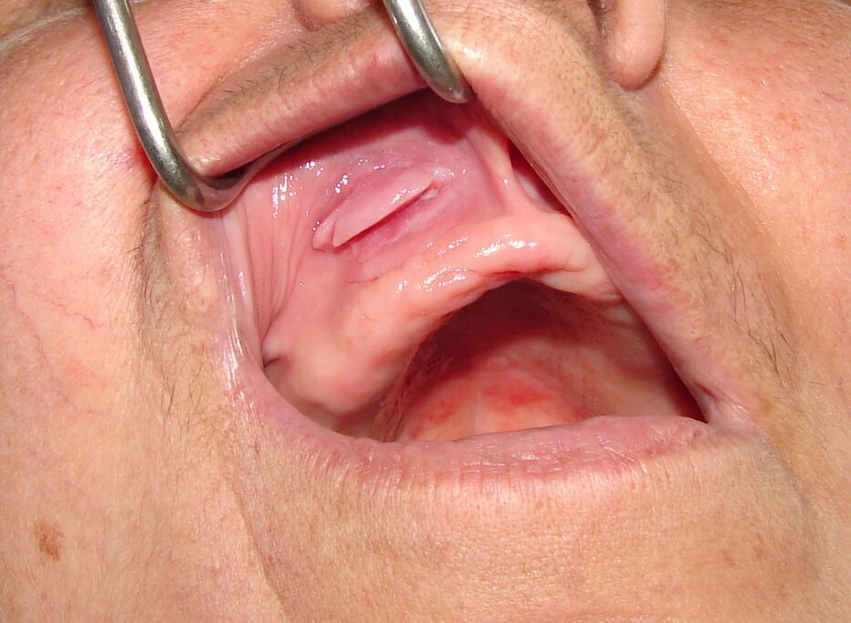
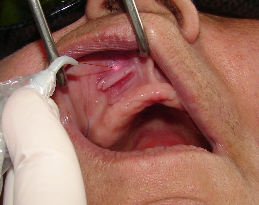
| Figure 1. The maxillary epulis fissuratum is extended to the midline causing problematic insertion of full denture. Figure 2. The fiber-optic of diode laser applied on the labial aspect area first, using specific pain-free parameters. | |
A 70-year-old female was referred by her prosthodontist for assessment of an upper anterior mucogingival overgrowth (Figure 1). She had an uneventful medical history. The recent dental history was composed of maxillary full dental removal and full denture restoration. No allergies were reported. The patient’s weight was 75 Kg; her height was 169 cm.
Clinical examination revealed the presence of epulis fissuratum. As a consequence, this presented pathological mechanical pressure on the associated acrylic flange of the full denture.
It was decided to perform laser excision of the pathological tissue without infiltrated anesthesia using specific laser parameters (Figure 2). The laser equipment was defined by the manufacturer (Lambda Scientifica Srl) as a class-II B device according to the CE conformity statement; the parameters were 1100 mW at 808 nm with continuous output. The optical fiber used was 300 μm in diameter, allowing a very fine soft tissue cut. According to the manufacturer, the optical protective glasses had an Optical Density>5 at the wavelength of emission from the diode. According to standard EN 60825 CEI 76-2 II, the minimum optical density has been estimated to be 4.96 at 0.05 m.
Pre-operatively, informed counseled consent was obtained both verbally and in written format on a consent form.
 |
| Figure 3 |
|---|
| Figure 3. The final elliptical laser cut did not require sutures or periodontal dressing. Postoperative hemostasis is optimum. |
The epulis was sprayed with lidocaine four times in 1-minute intervals. The laser fiber was applied almost vertically and anteriorly to the junction with the healthy mucosa, initially causing disruption of the mucosa continuity. This facilitated performing deeper laser assisted excision of the epulis in a horizontal dimension. The design of the excision was elliptical allowing complete removal of the epulis by using the laser fiber. The whole procedure was carried out within 10 minutes, without pain. No sutures were required. Hemostasis was achieved immediately after the procedure (Figure 3). The patient was comfortable with no pain, either intra-operatively or post-operatively. The patient described the procedure as totally painless. Ten days later healing was found to be uneventful. Histopathology revealed the benign nature of the condition.
Discussion
Epulis fissuratum is a benign condition mainly seen in geriatric patients. The presence of a denture is the nearly universal cause of this benign pathological condition. Dentures are removable prosthetic dental solutions, which means that there is always some micromovement during mastication. If the denture is ill fitting, the movement is excessive causing trauma to soft tissues, especially in non-keratinized oral mucosa.
The chronic oral trauma caused by sharp edges of teeth and ill-fitting dentures has been incriminated in the etiology of some oral cancers [1]. A recent study showed that chronic trauma of the oral mucosa is an important risk factor in patients with an oral cancer diagnosis but not so important in patients with potentially malignant oral disorders [2].
Initially, the erosion or inflammation of the mucosa may be caused by the acute irritation of the denture acrylic flange in the vestibular region. This is painful and shows all the signs and symptoms of acute inflammation. If the lesion remains untreated, the chronic consequence of this type of injury is clinically seen as fissured hyperplasia, epulis fissuratum. Sometimes, the lesion appears erosive related to an acute exacerbation of symptoms.
Treatment of epulis fissuratum may be conservative or surgical. A conservative approach should be considered as the first option because of its noninvasive nature. However, a conservative approach is time consuming and initially requires removal of the acrylic flange associated with the trauma and relining or repairing of the full denture. After a few weeks, when the lesion is completely healed, the acrylic flange may be relined and redesigned correctly to avoid further trauma of the mucosa when it is fitted in the mouth.
Surgical methods mainly include removal of the lesion using blade, electrocauterization, or laser. Blade excision always requires infiltrated or general anesthesia and sutures or periodontal dressing. The necessity of infiltrated or general anesthetic is essential in electrosurgery but usually without sutures or periodontal dressing.
The role of laser in dentistry is well-established in conservative management of oral diseases [3]. In oral surgery, it is still under evaluation [4, 5, 6]. This seems to be unavoidable if we consider that in surgical science it is difficult to perform an organized double-blind randomized controlled trial, which is a prerequisite for the evaluation of each surgical technique.
The diode light equipment may be considered a modern laser technology in the field of dentistry. Diode laser showed good results as an extra adjunct to the classical methods in the management of inflamed periodontal tissues and endodontics [5, 7, 8].
Currently, painless procedures are secured by using local or general anesthesia. This case report describes the parameters of performing maxillary removal of epulis fissuratum in a geriatric patient without infiltrated local anesthesia. Moreover, the reassurance of the patient about the painless procedure is one of the most important criteria.
Excision of epulis fissuratum is a common procedure in the field of oral and maxillofacial surgery. The advantage of laser surgery includes higher precision when compared to other surgical tools, which results in less pain, bleeding, swelling, and scarring. The procedure is not time consuming, easy to perform in an outpatient setting, and requires no sutures. All of these features combine to decrease the risk of post-operative infection [9].
Laser transmits energy to the cells causing warming, welding, coagulation, protein denaturization, drying, vaporization, and carbonization [10]. The great advantage of diode laser removal of epulis in geriatric patients should be the avoidance of needle-infiltrated anesthesia.
Another advantage is the reduced time required for epulis excision by using diode laser in pain-free parameters compared to electrosurgery and blade incision, which always require anesthesia. In severe cases of extended epulis the need for anesthesia is essential. Formal randomized controlled trials are required to decide which procedure is medically superior to others. We posit the use of a diode laser.
Pain is an unpleasant sensory and emotional experience associated with actual or potential tissue damage [11]. Currently, we suggest that a way of avoiding such an experience in gerodontics is to perform laser epulis excision without infiltrated anesthesia. In summary, pain is an objective feeling that is very difficult to assess. Pain perception is another important issue in creating management pathways for surgical problems. According to our experience every patient has different pain thresholds that are not age dependent. Therefore, we suggest initiating soft tissue laser treatment without infiltrated anesthesia at less than 1000 mW and if there is no pain, to increase power slowly until reaching the pain threshold. The need for a randomized controlled trial is emphasized in order to establish the exact efficacy of this technique when compared to other methods. It is obvious that diode laser epulis fissuratum excision may be performed without infiltrated anesthesia with optimum post-surgical healing. In severe cases of soft tissue excision the need for anesthesia may be essential [12]. Pain-free surgery is the aim in all types of surgical management; we have shown that this can now be achieved for selected conditions related to epulis fissuratum [6].
References
1. Rosenquist K. Risk factors in oral and oropharyngeal squamous cell carcinoma: a population-based case-control study in southern Sweden. Swed Dent J Suppl 2005;179:1-66. [PubMed]2. Piemonte ED, Lazos JP, Brunotto M. Relationship between chronic trauma of the oral mucosa, oral potentially malignant disorders and oral cancer. J Oral Pathol 2010 (Epub Ahead of Print). [PubMed]
3. Ishikawa I, Aoki A, Takasaki AA. Clinical application of erbium: YAG laser in periodontolgy. J Int Acad Periodontol 2008;10:22-30. [PubMed]
4. Kafas P, Kalfas S. Carbonization of a radicular cyst using fiber-optic diode laser: a case report. Cases J 2008;1:113. [PubMed]
5. Capodiferro S, Maiorano E, Scarpelli F, Favia G. Fibrolipoma of the lip treated by diode laser surgery: A case report. J Med Case Reports 2008;2:301. [PubMed]
6. Kafas P, Stavrianos C, Jerjes W, Upile T, Vourvachis M, Theodoridis M, Stavrianou I. Upper-lip laser frenectomy without infiltrated anaesthesia in a paediatric patient: a case report. Cases J 2009;2:7138. [PubMed]
7. de Souza EB, Cai S, Simionato MR, Lage-Marques JL. High-power diode laser in the disinfection in depth of the root canal dentin. Oral Surg Oral Med Oral Pathol Oral Radiol Endod 2008;106:e68-72. [PubMed]
8. Kafas P, Dabarakis N, Theodoridis M. Performing gingivoplasty without injected anaesthesia: a case report. Surg J 2008;3:27-29.
9. Kafas P, Angouridakis N, Dabarakis N, Jerjes W. Diode laser lingual frenectomy may be performed without local anaesthesia. Int J Orofac Sci 2008;1:1.
10. Sarver DM, Yanosky M. Principles of cosmetic dentistry in orthodontics: part 2. Soft tissue laser technology and cosmetic gingival contouring. Am J Orthod Dentofacial Orthop 2005;127:85-90. [PubMed]
11. Pain terms: a list with definitions and notes on usage. Recommended by the IASP Subcommittee on Taxonomy. Pain 1979;6:249. [PubMed]
12. Kato J, Wijeyeweera RL. The effect of CO(2) laser irradiation on oral soft tissue problems in children in Sri Lanka. Photomed Laser Surg 2007;25:264-268. [PubMed]
© 2010 Dermatology Online Journal